Positron emission tomography PET is a technique that measures physiological function by looking at blood flow metabolism neurotransmitters and radiolabelled drugs. Positron emission tomography PET scans produce detailed 3-dimensional images of the inside of the body.

A brain positron emission tomography PET scan is an imaging test that allows doctors to see how your brain is functioning.
Positron emission tomography how it works. Positron emission tomography PET is a type of nuclear medicine procedure that measures metabolic activity of the cells of body tissues. PET is actually a combination of nuclear medicine and biochemical analysis. Used mostly in patients with brain or heart conditions and cancer PET helps to visualize the biochemical changes taking place in the body such as the metabolism the process by which cells.
Posted March 23 2016 anosh01. This post aims to explain what positron emission tomography PET is and how it works. PET is a unique type of medical imaging that reveals information about the physiology of organs and tissues unlike CT or MRI machines which only yield images of anatomy.
By doing this PET scans can often detect irregularities such as cancer significantly earlier than other diagnostic tests. A positron emission tomography PET scan is an imaging test that allows your doctor to check for diseases in your body. The scan uses a.
How do they workTo find o. Positron Emission Tomography PET scans are a way of imaging body functions in 3D using specially designed radioactive molecules. A positron emission tomography also known as a PET scan uses radiation to show activity within the body on a cellular level.
It is most commonly used in. Because positron emission tomography is a mouthful radiologists call it a PET scan for short. Doctors often use the diagnostic exam often to detect cancer and measure the effects of cancer.
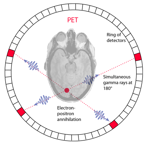
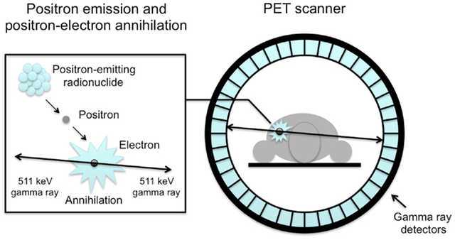
Positron Emission Tomography PET Figure 2 PET produces images of the body by detecting the radiation emitted from radioactive substances. These substances are injected into the body and are usually tagged with a radioactive atom such as Carbon-11 Fluorine-18 Oxygen-15 or Nitrogen-13 that has a short decay time. Even with newer and repurposed anti-TB drugs almost a third of patients with XDR-TB have unfavourable outcomes.
In patients with localised disease and adequate pulmonary reserve surgery is an important adjunctive treatment. However there are no outcome data from TB endemic countries and the prognostic significance of pre-operative PET-CT findings remains. Positron emission tomography PET scans produce detailed 3-dimensional images of the inside of the body.
The images can clearly show the part of the body being investigated including any abnormal areas and can highlight how well certain functions of the body are working. PET scans are often combined with CT scans to produce even more detailed images. Positron emission tomography also known as PET scan is a medical imaging approach that utilizes a radioactive elementdrug to analyze the organ and tissue functionality and is well-known for its.
Positron emission tomography PET is a technique that measures physiological function by looking at blood flow metabolism neurotransmitters and radiolabelled drugs. PET offers quantitative analyses allowing relative changes over time to be monitored as a disease process evolves or in response to a specific stimulus. Positron emission tomography PET is a technique that measures physiological function by looking at blood flow metabolism neurotransmitters and radiolabelled drugs.
PET offers quantitative analyses allowing relative changes over time to be monitored as a disease process evolves or in response to a specific stimulus. How Positron Emission Tomography Works. In PET scanning the engine detects radiation emitted by the radiotracer.
Radiotracer is made up of radioactive materials characterized by natural chemicals such as glucose. The radiotracer is injected into the body where it moves into the cells that use glucose for energy. The goal of the video youre about to see is to describe the procedure for conducting nuclear medicine tests using positron emission tomography commonly cal.
A brain positron emission tomography PET scan is an imaging test that allows doctors to see how your brain is functioning. The scan captures images of the activity of the brain after radioactive. Positron emission tomography PET uses small amounts of radioactive materials called radiotracers or radiopharmaceuticals a special camera and a computer to evaluate organ and tissue functions.
By identifying changes at the cellular level PET may detect the early onset of. How does Positron Emission Tomography PET work. This is a complex topic but Ill try to put in lay terms.
PET scanning uses elements of computer aided cross sectional imaging similar to CT and MRI scanning and nuclear medicine imaging.