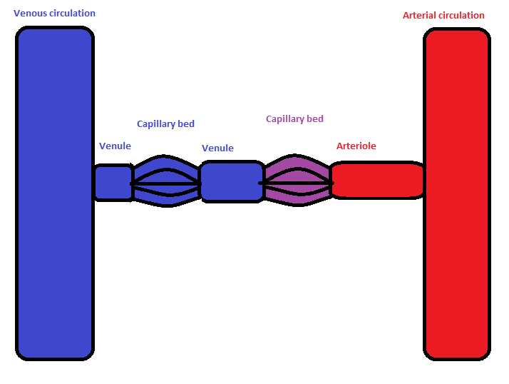
Examples of such systems include the hepatic portal system the hypophyseal portal system and in non-mammals the renal portal system. Portal venous systems are considered venous because the blood vessels that join the two capillary beds are either veins or venules.

Ultrasound Evaluation of the Portal and Hepatic Veins Leslie M.
Portal venous system images. Portal vein thrombosis illustration - portal vein stock pictures royalty-free photos images human blood circulation lithograph published in 1882 - portal vein stock illustrations Illustration from Surgical Anatomy. The Treatise of the Human Anatomy and Its Applications to the Practice of Medicine and Surgery volume III. Human anatomy scientific illustrations.
Portal venous system - hepatic portal system stock illustrations Blood Circulation Illustration Circulatory System Vein-Artery-Heart In. In the circulatory system of animals a portal venous system occurs when a capillary bed pools into another capillary bed through veins without first going through the heartBoth capillary beds and the blood vessels that connect them are considered part of the portal venous system. They are relatively uncommon as the majority of capillary beds drain into veins which then drain into the.
The portal venous system typically consists of the portal vein which is formed by the union of the splenic vein with the superior mesenteric vein and several levels of portal venous branches. It carries blood from the abdominal portion of the digestive system spleen pancreas and gallbladder to. A portal venous varix plural.
Portal venous varices refers to a segments of aneurysmal variceal dilatation of the portal vein are extremely rare and represent only 3 of all aneurysms of the venous systemThey are still however the most common visceral varix 8. Congenital anomalies of the portal venous system comprise total or partial agenesis of the portal vein abnormal branching of the portal vein venous malposition in situs inversus totalis or in midgut malrotation arteriovenous malformations and persistence of fetal valves. The portal vein is the primary collateral route for decompression of the liver in elevated pressure.
In a normal state the portal venous system is a low-pressure system with a normal pressure of 5 to 10 mm Hg. DOPPLER SPECTRAL ANALYSIS Portal and Hepatic System The portal vein normally exhibits a monophasic. Ultrasound Evaluation of the Portal and Hepatic Veins Leslie M.
Revzin Hjalti Thorisson Ulrike M. Hamper Portal hypertension PHT is an extremely common medical problem worldwide. In Western countries PHT most commonly occurs secondary to underlying liver cirrhosis either viral or alcohol induced.
Morbidity is primarily related to bleeding from. Portal venous gas is the accumulation of gas in the portal vein and its branches. It needs to be distinguished from pneumobilia although this is usually not too problematic when associated findings are taken into account along with the pattern of gas ie.
Peripheral in portal venous gas. Portal-to-systemic venous communications are frequently seen on imaging studies in patients with portal hypertension from cirrhosis. These communications are largely extrahepatic and are commonly via the coronary vein esophageal varices or retroperitoneal collaterals.
A portal venous system is a deviation from this configuration. It occurs when a capillary bed drains into another capillary bed before going back to the heart. Its a venous system because the vessels that connect the 2 capillary beds are veins.
They contain deoxygenated blood. Portal venous systems are considered venous because the blood vessels that join the two capillary beds are either veins or venules. Examples of such systems include the hepatic portal system the hypophyseal portal system and in non-mammals the renal portal system.
Unqualified portal venous system. Pneumobilia also known as aerobilia is the accumulation of gas in the biliary treeIt is important to distinguish pneumobilia from portal venous gas the other type of branching hepatic gasThere are many causes of pneumobilia and clinical context is often important to distinguish between these 3. Postprocessing techniques maximum intensity projection MIP multiplanar reconstruction or volume rendering were used for detailed analysis of portal venous system aneurysms or suspected portal venous system aneurysms.
The thickness of the MIP images used in the study ranged from 5 to 50 mm but most ranged from 10 to 25 mm. In volunteers and patients portal volumetric flow rates determined by using MR images and Doppler sonography showed good correlation r 94. Our results indicate that cine phase-contrast MR imaging is a practical noninvasive method for measuring volumetric flow rates in the portal venous system.