Ct is p. This technology involves several sets of CT scans taken over several minutes.

On the day of admission scoring systems based on imaging do not outperform scoring systems based on clinical and biochemical parameters with regard to predicting clinical outcome.
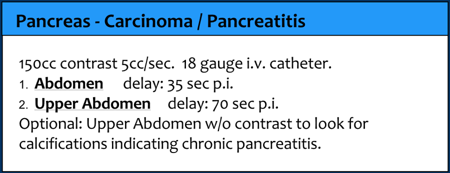
Pancreatic protocol ct scan. Pancreatic protocol CT involves imaging at the pancreatic phase ie approximately 45 seconds after contrast administration and at the portal venous phase ie approximately 70 seconds after contrast administration. It is useful for detection of adenocarcinoma of pancreas. These techniques are called pancreatic protocols.
This is the preferred CT scan type for diagnosing pancreatic cancer. If a pancreatic protocol CT is not available a high-quality CT scan may work. Another type is the combined positron emission tomography computed tomography PET-CT.
This is called a multi-phase CT scan or a pancreatic protocol CT scan. CT scans can give healthcare providers more information related to injuries or diseases of the pancreas. Why might I need a CT scan of the pancreas.
A CT scan of the pancreas may be used to check the pancreas for. Tumors or other lesions. CT examination of the pancreas should always be done with maximum amount of contrast at a maximum flow rate because both small pancreatic carcinomas aswell as pancreatic necrosis in pancreatitis are difficult to detect.
It is a matter of personal flavor to do the whole abdomen at 35 sec pi. Or at 70 sec pi. First timeinitial elevated lipase or rule-out pancreatitis routine AbdPel 70s single phase Chronic pancreatitis routine Abd 70s single phase Patient Position.
Supine feet down with arms above head Scan Range CC z-axis. 1 cm above diaphragm through superior iliac crest Prep. No solids liquids OK for 3 hours prior to examination.
Currently a typical pancreatic MDCT protocol is a dual-phase CT acquisition after IV contrast medium administration at a flow rate of 35 mLs for optimal pancreatic CT enhancement. The pancreatic phase is performed around 3540 seconds after contrast agent injection and the portal venous phase PVP acquisition is subsequently performed after a 70-second delay 19 20. The CT severity index CTSI combines the Balthazar grade 0-4 points with the extent of pancreatic necrosis 0-6 points on a 10-point severity scale.
On the day of admission scoring systems based on imaging do not outperform scoring systems based on clinical and biochemical parameters with regard to predicting clinical outcome. A pancreatic CT scan may diagnose injuries and diseases of the pancreas. CT scans of the pancreas can identify tumors masses or cysts on the pancreas or in the surrounding structures of the pancreas.
A CT scan of the pancreas may help diagnose pancreatic cancer and pancreatitis. What is a CT scan of the pancreas. Computed tomography CT scan is a noninvasive diagnostic imaging procedure that uses a combination of X-rays and computer technology to produce horizontal or axial images often called slices of the body.
A CT scan shows detailed images of any part of the body including the bones muscles fat and organs. This is called a multi-phase CT scan or a pancreatic protocol CT scan. CT scans can give healthcare providers more information related to injuries or diseases of the pancreas.
Why might I need a CT scan of the pancreas. A CT scan of the pancreas may be used to check the pancreas for. Tumors or other lesions.
Pancreatic CT protocol improves image quality reduces radiation dose Following a patient-specific contrast media protocol during CT of the pancreas can enhance image quality reduce contrast volume and reduce radiation dose according to a new study published in Academic Radiology. Multiphase multidetector computed tomography CT of the pancreas is widely used for screening detecting staging and following up pancreatic cancer The standard protocol used in our institution includes a low-milliamperage unenhanced CT scan of the abdomen followed by a late arterial pancreatic phase scan of the pancreas and a portal venous phase scan of the abdomen. CT will show both.
Yes a ct scan will show both organs the pancreas and the colon. Contrast material such as oral or IV contrast may enhance the evaluation. Ct is p.
Also known as pancreatic protocol CT this helps identify staging and whether or not surgery would be an effective option. This technology involves several sets of CT scans taken over several minutes. In short a CT computerized tomography scan can show the pancreas pretty clearly and is a trusted modality when pancreatic cancer is suspected.
A CT protocol is a set of parameters that specify a specific exam and contrast delivery requirements. When a CT scan is requested it will be vetted by a radiologist or radiographer to determine the study is justified and what the most suitable parameters by which that CT should be performed - this may lead to a different CT examination being performed or an alternative modality recommended. 111 For people with obstructive jaundice and suspected pancreatic cancer offer a pancreatic protocol CT scan before draining the bile duct.
112 If the diagnosis is still unclear offer fluorodeoxyglucose-positron emission tomographyCT FDGPETCT andor endoscopic ultrasound EUS with EUSguided tissue sampling.