This is calculated by 052 x length x width x height. Your doctor will typically use an endorectal coil with low-field 15 Tesla MRI magnets if you have a metal orthopedic implant.

Getting a detailed look at some pictures of prostate cancer can give you a better idea of what youre up against.
Normal prostate mri images. Normal volume of the prostate should be less than 30 ml. This is calculated by 052 x length x width x height. MRI imaging is helpful in differentiation the prostatic zonal anatomy best demonstrated on T2WI.
Normal volume of the prostate should be less than30 ml. This is calculated by 052 x length x width x height. MRI imaging is helpful in differentiation the prostatic zonal anatomy best demonstrated on T2WI.
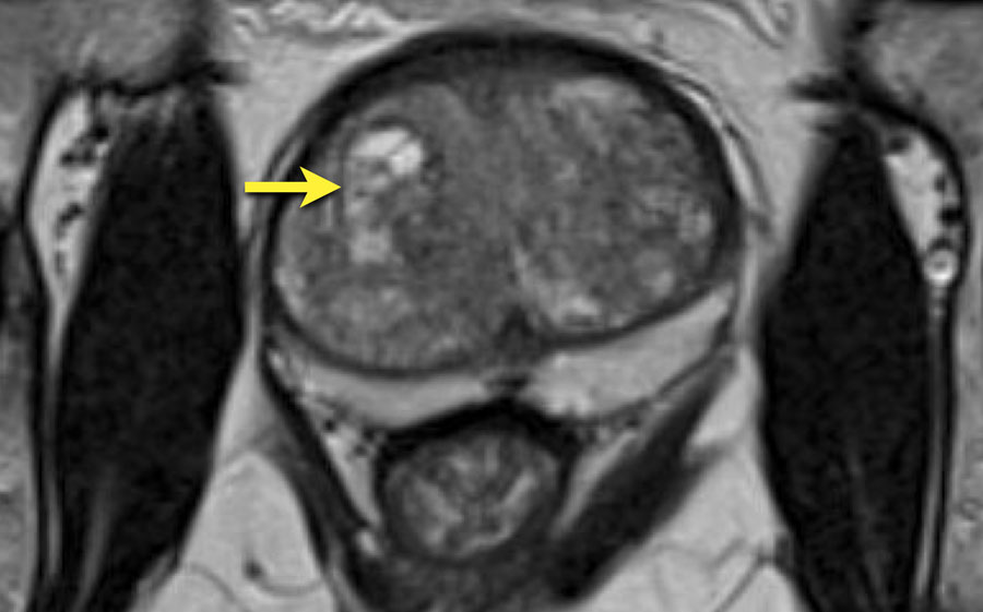
Normal prostate anatomy on MRI. Axial a b sagittal c and coronal d T2-weighted MR images of the male pelvis. The seminal vesicles arrows in a and d drain into the midprostatic urethra via the ejaculatory ducts arrow in c at the level of the verumontanum arrowhead in c.
With imagers operating at 035 and 15 T T2-based tissue-contrast images were needed for the demonstration of the internal anatomy of the prostate gland. The display of zonal anatomy was improved when continuous 05-cm slices were used. Evaluating sequential sections the peripheral central and transition zones could be differentiated.
A magnetic resonance imaging MRI scanner uses strong magnetic fields to create an image or picture of the prostate and surrounding tissues. The prostate gland is a small soft structure about the size and shape of a walnut which lies deep in the pelvis between the bladder and the penis and in front of the rectum back passage. Prostate MRI Prostate MRI Basic facts about PCA How When MRI Technique Why do MRI and What to Look for Prostate Cancer PSA level Gleason Score 3 4 4 3 T Stage T2c T3a T3b.
1162010 2 Axial T2 sagT2 right SV sagT2 left SV Staging PPA coil ERC-PPA coil Value of T1-weighted Images Why Main Role of MRI Local Staging of Prostate Cancer Extracapular. Field of view FOV should encompass the entire prostate gland and seminal vesicles and it generally ranges between 12 and 20cm. Diffusion-weighted imaging DWI which reflects the random motion of water molecules and is a key component of the prostate mpMRI exam but it must be correlated with T2W images as it very poorly reflects anatomy.
Getting a detailed look at some pictures of prostate cancer can give you a better idea of what youre up against. A number of prostate cancer images have been compiled to aid in your education. A rectal exam is a common procedure your doctor may perform to look for prostate cancer warning signs.
Additionally prostate MRI may examine water molecule motion water diffusion and blood flow perfusion imaging within the prostate to help differentiate between diseased and normal prostate tissue. Your doctor will typically use an endorectal coil with low-field 15 Tesla MRI magnets if you have a metal orthopedic implant. MRI TECHNIQUES FOR PROSTATE CANCER EVALUATION Conventional MRI T2 T1 Optimal MRI for prostate cancer detection requires the use of an endorectal coil and a pelvic phased-array coil with a magnet of 15 Tesla T or higher thin 3-mm slices and a small 14-cm field of view 1.
On T2-weighted MR images the zonal anatomy of the prostate. Multiparametric MRI evaluation of the prostate includes three general components. High-resolution T2WI and at least two functional MRI techniques including diffusion weighted imaging and either MR spectroscopic imaging MRSI or dynamic contrast enhanced MRI DCE-MRI.
T2WI provides the best depiction of the prostates zonal anatomy and capsule. The scan shown demonstrates the normal zonal anatomy of the prostate. The prostate gland can be viewed as having 4 major zones.
In the normal gland the outer zone called the peripheral zone accounts for 70 of the parenchyma. It is often canoe shaped in the axial projection. Normal prostate anatomy on MRI.
The normal prostate gland demonstrates intermediate to low signal intensity on T1-weighted imaging 4. There is insufficient soft-tissue contrast resolution on the T1W images to distinguish the intraglandular architecture and therefore this sequence is not used for tumour localisation Figure 1. Axial T1 image showing homogeneous low signal.
Prior to biopsy for prostate cancer diagnosis Post radiotherapy and chemotherapy assessment Infection prostatitis or prostate abscess Newly diagnosed prostate cancer staging Diagnosis of recurrent prostate cancer Post prostatectomy assessment Pre surgical assessment Tumor detection and staging Congenital abnormalities. If you have a raised PSA level your doctor may refer you to hospital for an MRI scan of your prostate. If the scan shows a problem it can be targeted later with a biopsy.
Having a biopsy to diagnose prostate cancer. There are a few types of biopsy that may be used in hospital including the below. A transperineal biopsy.
This is where a needle is inserted into the prostate through.