Two independent observers evaluated the following features on the randomized CT studies. Suboptimal portal vein blood flow can be difficult to distinguish from suboptimal hepatic venous drainage.

File name man002jpg Description title page.
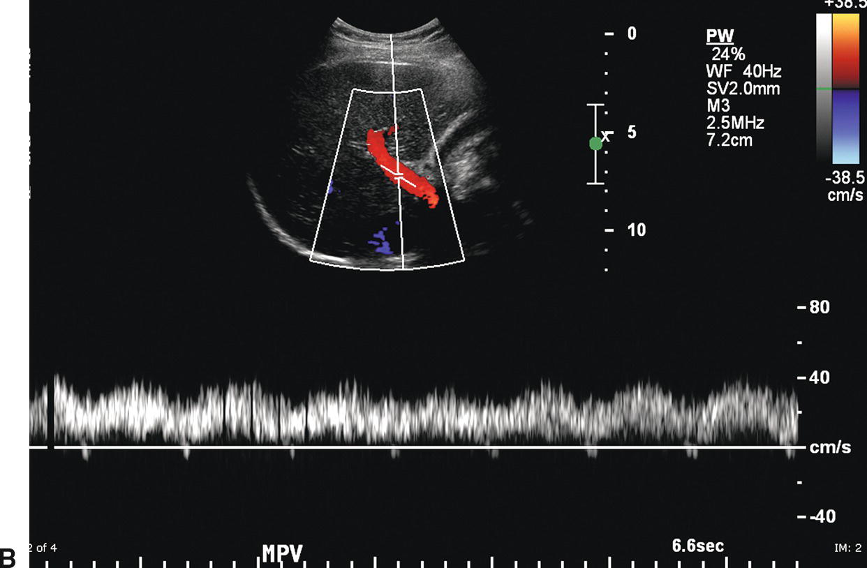
Hepatopetal flow within the portal vein. Hepatopetal denotes flow of blood towards the liver which is the normal direction of blood flow through the portal vein. The term is typically used when discussing the portal vein or recanalized vein of the ligamentum teres in patients with suspected portal hypertension. It is the opposite of hepatofugal.
Hepatopetal flow within the portal vein. They have highly echogenic walls they course horizontally in the liver they are inferior to hepatic veins they have hepatopetal flow their diameter decreases as they travel within the liver and they are intrasegmental. Normally the portal vein receives flow from the superior mesenteric vein and the splenic vein.
Flow is hepatopetal mean Answered by. Shunted hepatic artery blood striped red arrow has precipitated hepatofugal flow in the portal vein branch that normally supplies the region where the lesion is situated. The shunted blood then joins normal hepatope-tal blood flow in an adjacent portal vein branch solid red arrow.
Blue arrows normal portal vein flow green ar-. The normal portal vein measures less than 13 mm during quiet respiration. Color Doppler interrogation revealed no evidence of thrombus.
MPV main portal vein LPV left portal vein. B Spectral Doppler waveform from the portal vein demonstrating decreased velocity. Flow remains hepatopetal heading toward the liver.
Hepatofugal or non-forward portal flow NFPF is an abnormal flow pattern in which the portal venous flow is from the periphery of the liver towards the porta and backwards along the portal vein. This phenomenon is not uncommon in patients with liver disease 3. Hepatofugal flow is typically seen in the right portal vein due to the sump effect of the paraumbilical vein.
The left portal vein remains hepatopetal but may become enlarged as it feeds the paraumbilical vein. Portal vein is the main blood flow to the liver it occluded sometime with thrombosis that is usually treated with blood thinners like Coumadin warfarin. Hepatic veins drains blood from liver to the heart if they occlude the patient will develop jaundice and sometimes severe liver dysfunction.
This week Julie discusses flow direction and characteristics of the portal and the hepatic veins. In most system settings a normal main portal vein is displ. Suboptimal portal vein blood flow can be difficult to distinguish from suboptimal hepatic venous drainage.
Linear zones of ischemic necrosis andor hepatocellular atrophy favor the former whereas red blood cell congestion within central veins and centrilobular sinusoids and obliterative central venopathy favor the latter. Ultrasonography andor angiography are needed to more accurately characterize the cause of the vascular abnormality. Cases of suboptimal portal vein flow.
Hepatofugal flow ie flow directed away from the liver is abnormal in any segment of the portal venous system and is more common than previously believed. Hepatofugal flow can be demonstrated at. A Complete hepatofugal flow in intrahepatic portal vessels and main portal vein through splenic vein and splenorenal shunts.
B Complete hepatofugal flow in intrahepatic portal vessels and main portal vein through a dilated left gastric vein. C RF only in one or more intrahepatic portal vessels together with hepatopetal flow in main portal vein. The mechanism of reversal of flow in intrahepatic.
Normally the portal vein receives flow from the superior mesenteric vein and the splenic vein. In patients with cirrhosis and hepatofugal flow in the main portal vein the portal vein is supplied only by the hepatic artery which also supplies the hepatic veins. This decrease in flow volume could explain the decreased diameter of the portal vein.
Sis 18 with hepatopetal and 18 with hepatofugal flow in the main portal vein who underwent contemporaneous abdominal sonography and CT. Two independent observers evaluated the following features on the randomized CT studies. Diameter of the portal splenic and superior.
Flow is in the opposite direction red in the hepatic artery which lies anterior to the main portal vein. Reversed flow in the portal vein usually indicates relatively severe PHT and is a late finding. Doppler findings in the hepatic artery.
Congenital absence of the portal vein CAPV known as Abernethy malformation is a rare malformation and is more common in females. We present a case of a woman with CAPV and a coexisting intrahepatic inferior vena cava IVC branch showing hepatopetal flow which to our knowledge has not been reported in the medical literature. File name man001jpg Description front cover.
File name man002jpg Description title page. File name man003jpg Description copyright statement.