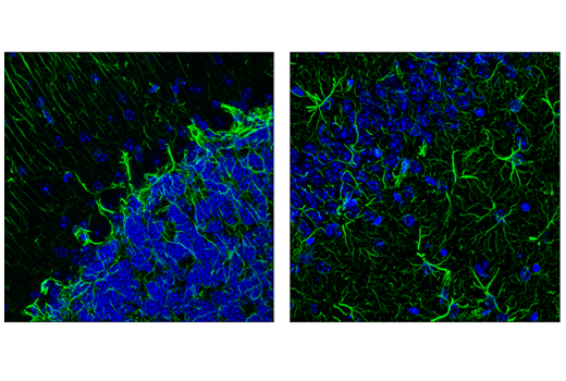
Our findings suggest that LIF signaling induces GFAP progenitor cells whereas BMP signaling. Whether LIF and BMP signaling generate GFAP-expressing cells with similar characteristics and developmental potential.

CST expects that GFAP GA5 Mouse mAb Alexa Fluor 488 Conjugate will also recognize GFAP in these species.
Gfap antibody cell signaling. This Cell Signaling Technology antibody is conjugated to Alexa Fluor 488 fluorescent dye and tested in-house for direct immunofluorescence of rat cells. The unconjugated antibody GFAP GA5 Mouse mAb 3670 reacts with human mouse and rat GFAP protein. CST expects that GFAP GA5 Mouse mAb Alexa Fluor 488 Conjugate will also recognize GFAP in these species.
The GFAP GA5 Mouse mAb Antibody from Cell Signaling Technology is a Mouse Monoclonal antibody to GA5 to Glial Fibrillary Acidic Protein and GFAP. GFAP GA5 Mouse mAb detects endogenous levels of total GFAP protein. Antibodies to GFAP are very useful as markers of astrocytic cells.
In addition many types of brain tumor presumably derived from astrocytic cells heavily express GFAP. GFAP is also found in the lens epithelium Kupffer cells of the liver in some cells in salivary tumors and has been reported in erythrocytes. GFAP is used as a marker to distinguish astrocytes from other glial cells during.
GFP Antibody detects GFP YFP and CFP-tagged proteins exogenously expressed in cells. This antibody does not detect RFP-tagged proteins. Please note that the GFP YFP and CFP tags add approximately 27 kDa to the molecular weight of the fusion protein.
Wash sections in wash buffer for 5 min. Block each section with 100400 µl of preferred blocking solution for 1 hr at room temperature. Remove blocking solution and add 100400 µl primary antibody diluted in SignalStain Antibody Diluent 8112 to each section.
Incubate overnight at 4C. GFAP is highly expressed in the central nervous system almost exclusively in astrocytes. It is the main filament protein of mature astrocytes and so seems to play a role in modulating astrocyte motility and shape1 As an intracellular antigen antibodies against GFAP are probably non-pathogenic but serve as a marker of cytotoxic T-cell-mediated autoimmunity2 3 Fang et al1 first reported.
Gfap 和波形纤维蛋白形成星形胶质细胞中的中间纤丝并且调节其运动性和形状 1具体而言波形纤维蛋白纤丝在发育早期阶段存在而 gfap 纤丝是分化和成熟的脑星形细胞的特征因此gfap 常用作源自星形细胞的颅内和椎管内肿瘤的标记物 2此外gfap 中间纤维也存在于周围神经系统内非. This Cell Signaling Technology antibody is conjugated to Alexa Fluor 555 fluorescent dye and tested in-house for direct immunofluorescence of rat cerebellum. The unconjugated antibody 3670 reacts with human mouse and rat GFAP protein.
CST expects that GFAP GA5 Mouse mAb Alexa Fluor 555 Conjugate will also recognize GFAP in these species. ALXDRDFLJ45472GFAPGlial fibrillary acidic protein Immunogen. Monoclonal antibody is produced by immunizing animals with native GFAP purified from pig spinal cord.
Glial fibrillary acidic protein GFAP_HUMAN. B2RD44 D3DX59 E9PAX3 P14136 Q53H98 Q5D055 Q6ZQS3 Q7Z5J6 Q7Z5J7 Q96KS4 Q96P18 Q9UFD0. Anti-GFAP Antibody Products from Cell Signaling Technology.
Anti-GFAP antibodies are offered by a number of suppliers. This target gene encodes the protein glial fibrillary acidic protein in humans and may also be known as ALXDRD. Structurally the protein is reported to be 499 kilodaltons in mass.
Based on gene name canine porcine monkey mouse and rat orthologs may also be found. Established in Beverly MA in 1999 Cell Signaling Technology CST is a privately-owned company with over 400 employees worldwide. We are dedicated to providing innovative research tools that are used to help define mechanisms underlying cell function and disease.
Since its inception CST has become the world leader in the production of the highest quality activation-state and total protein antibodies utilized to expand knowledge of cell signaling. ALXDRDFLJ45472GFAPGlial fibrillary acidic protein Immunogen. Monoclonal antibody is produced by immunizing animals with a synthetic peptide corresponding to residues surrounding Asp395 of human GFAP protein.
Glial fibrillary acidic protein GFAP. GFAP is involved in intracellular cytoskeletal reorganization cell adhesion maintenance of myelination and neuronal structure in the brain involvement in the formation of the blood-brain barrier and regulation of synapses. It is also involved in cell migration movement and mitosis.
Whether LIF and BMP signaling generate GFAP-expressing cells with similar characteristics and developmental potential. We therefore used a combined in vitro and in vivo approach to compare the properties of GFAP-expressing cells that are generated in response to LIF versus BMP signaling. Our findings suggest that LIF signaling induces GFAP progenitor cells whereas BMP signaling.
Immunocytochemistry - Anti-GFAP antibody ab4674 ab4674 staining GFAP in primary hippocampal rat neuronsglia obtained from Neuromics cat. The cells were fixed with 100 methanol 5 min permeabilized with 01 PBS-Tween for 5 minutes and then blocked with 1 BSA10 normal goat serum03M glycine in 01PBS-Tween for 1h.