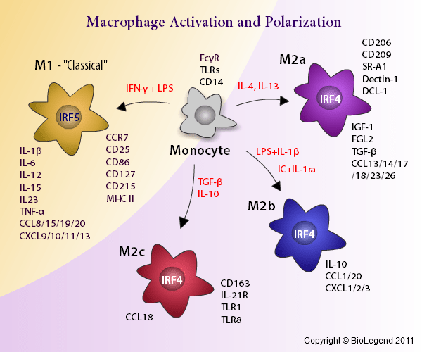
The mannose receptor CD206 a prototypic mouse M2 marker is highly expressed in M-CSFtreated macrophages in mouse whereas in human monocytes CD206 is substantially induced by GM-CSF 9 10. The images were taken under original magnification of 200.

1 M2 macrophages have numerous functions that can promote the proliferation of tumor cells and metastasis.
Cd206 m2 macrophage marker. When analyzed in a meta-analysis CD206 as a continuous variable showed superior predictive performance than classical prognosticators in AML BAALC ERG EVI1 MN1 and WT1. In summary M2 macrophages are preferentially enriched in AML. The M2 marker CD206 may serve as a new prognostic marker in AML.
Additionally a decrease in the M1 surface marker CD86 and an increase in the M2 surface marker CD206 were observed which suggested that M1 macrophages were polarized to an M2-like phenotype. Conditioned medium from these M2-like macrophages presented lower levels of proinflammatory cytokines and higher levels of proangiogenic factors and promoted endothelial cell proliferation. Macrophage Activation Markers CD163 and CD206 in Acute-on-Chronic Liver Failure.
Macrophages facilitate essential homeostatic functions eg endocytosis phagocytosis and signaling during inflammation and express a variety of scavenger receptors including CD163 and CD206 which are upregulated in response to inflammation. C AZM significantly increases the proportion of macrophages positive for the M2 marker CD206 and Arg-1 at 3 dpi but not 7 dpi relative to vehicle. E Representative images of CD206.
CD206 is a C-type lectin alternatively termed as the macrophage mannose receptor that is generally expressed by tissue macrophages dendritic cells and specific lymphatic or endothelial cells. M2 macrophages have anti-inflammatory and tissue repair functions and are characterised by CD36 CD206 and CD163 expression 8910. This model is simplistic as macrophages.
Likewise known M2 macrophage markers Arg1 Chi3l3Ym1 RetnlaFizz1 Egr2 Fn1 and Mrc1CD206 were also expressed in our M2 dataset labeled in Fig 1B. These data confirm those described in previous mouse or human M1 or M2 macrophage analyses and validate the robustness of our array results for more detailed data mining. Inducible nitric oxide synthase iNOS an important marker of M1 macrophage activation is located in the cytoplasm.
CD206 a highly expressed cell-surface protein on M2 macrophages is a marker for M2 polarization 32 33. Therefore iNOS and CD206. Both M1 and M2 macrophages expressed similar level of CD86CD80 markers of M1 macrophage and CD206 marker of M2 macrophage in all groups of CAD patient Fig.
M2 macrophages cultured from obstructive CAD patients have significantly higher level of CD11b CD14 and most importantly CD200R as compared to M1 macrophages. Our findings indicate that CD206 M2-like macrophages in adipose tissues create a microenvironment that inhibits growth and differentiation of adipocyte progenitors and. The mannose receptor CD206 a prototypic mouse M2 marker is highly expressed in M-CSFtreated macrophages in mouse whereas in human monocytes CD206 is substantially induced by GM-CSF 9 10.
CD163 scavenger-R type A another mouse M2 marker is highly expressed after IL-10 treatment in human but not after IL-4 treatment. Additionally the M1 phenotype transformation of macrophages was evidenced by the increased expression levels of CD86 tumor necrosis factor TNFα and inducible nitric oxide synthase iNOS and by the decreased expression levels of CD206 Sirt6 interleukin IL4 and IL10. The images were taken under original magnification of 200.
F Representative results for coimmunostaining of CD68 a macrophage marker and CD206 an M2 marker in the lung sections from patients with COVID-19. The CD163 CD204 and CD206 antigens are known to be upregulated in M2-type macrophages and are considered to be M2 markers. CD163 and CD204 macrophage scavenger receptor class A are receptors of the hemoglobinhaptoglobin complex and modified LDL respectively although recent studies have demonstrated that these receptors bind many ligands such as bacteria.
Tumor associated macrophages can compose up to half of the cell mass within a breast tumor. 1 M2 macrophages have numerous functions that can promote the proliferation of tumor cells and metastasis. 1 This study investigated CD206 and CD68 expression macrophage markers in the tumor stroma as a prognostic factor for overall survival and risk of recurrence in patients with locally advanced breast.
As CD206 is a M2 MΦ specific marker CD206 DTR mice which was recently established in our institute 2627 would be useful to investigate the role of M2 MΦs in folliculogenesis. Macrophages MΦs are involved in folliculogenesis and ovulation. However it is unknown which type of MΦ M1 or M2 plays a more essential role in the ovary.
CD206 or CD11c diphtheria toxin receptor transgenic DTR mice which enable depletion of CD206 M2 MΦs and CD11c MΦ or CD11c Dendritic cells DCs respectively were used. Background Alternatively activated M2 macrophages are phenotypically characterized by the expression of specific markers mainly macrophage scavenger receptors CD204 and CD163 and mannose receptor-1 CD206 and participate in the fibrotic process by over-producing pro-fibrotic molecules such as transforming growth factor-beta1 TGFbeta1 and metalloproteinase MMP-9.